
introductory
Preterm birth affects approximately 10% newborns globally and is considered to be the most important risk factor for impaired neurocognitive development, in addition, preterm infants may be at a 7-fold higher risk of adverse neonatal outcomes than normal children (Johnson and Marlow, 2016) Preterm birth has a significant impact on motor development in addition to neurocognitive development (Hadders-Algra, 2018). The main treatments used to promote normal motor development in children are reflexology and massage. Both techniques have been shown to be feasible and safe; however, the mechanism of action at the brain level is unknown (Beetz et al., 1983). In this study, experiments and data were analyzed in normal and preterm infants using the technique of simultaneous EEG and NIR multimodal acquisition.
01
Experimental background
Measuring brain activity and oxygenation in infants relies primarily on EEG and fNIRS technologies. eEG records electrical activity in the brain through scalp electrodes and is used to assess neonatal conditions and therapeutic effects, and is particularly suited to detecting epilepsy and neurological disorders. fNIRS measures changes in cerebral oxygenation through near-infrared light, which is useful for studying brain function and oxygenation in infants and children.
With increasing gestational age, EEG activity in preterm infants gradually becomes continuous and rhythmic from intermittent high-amplitude δ-wave bursts. Preterm birth is a major risk factor for neurocognitive development in infants and may lead to motor development problems.
Early interventions, such as Vojta Reflex Movement Therapy, may help promote neuroplasticity and normal motor development in these infants.
02
Experimental Methods
01
subject's choice
Inclusion criteria:Clinically stable preterm births between 1 and 3 months of age (
Exclusion Criteria:The presence of a neurological or respiratory disease in the newborn, an accident or complication during delivery, or being or having been treated for any disease.
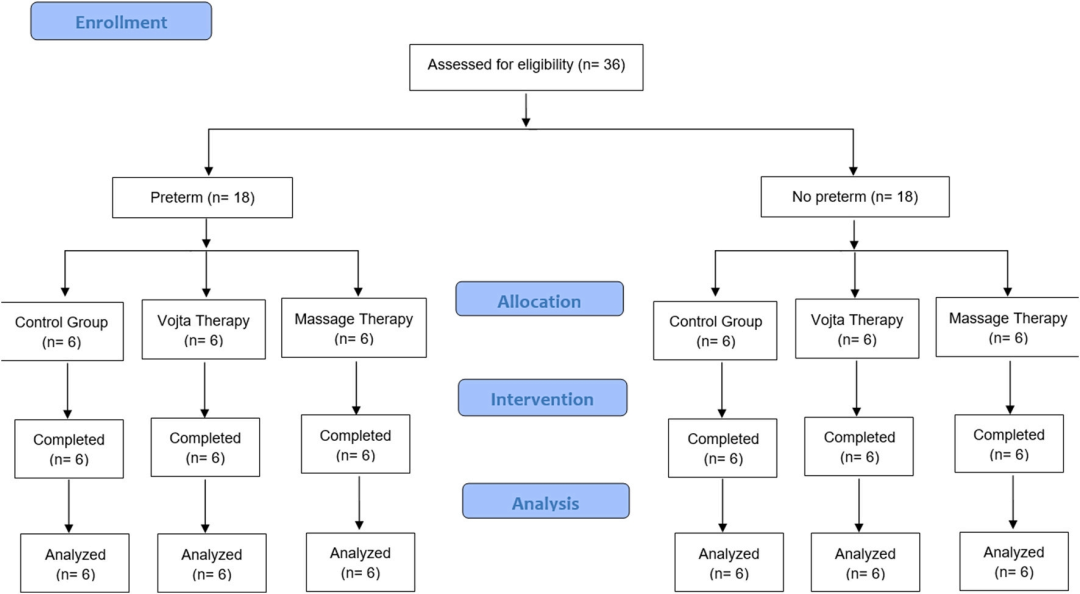
Sample size calculation:A mixed ANOVA analysis was performed using the G - Power program to determine a sample size of 6 subjects per group for the pilot study and to calculate the required sample size.
02
Experimental design


03
data acquisition
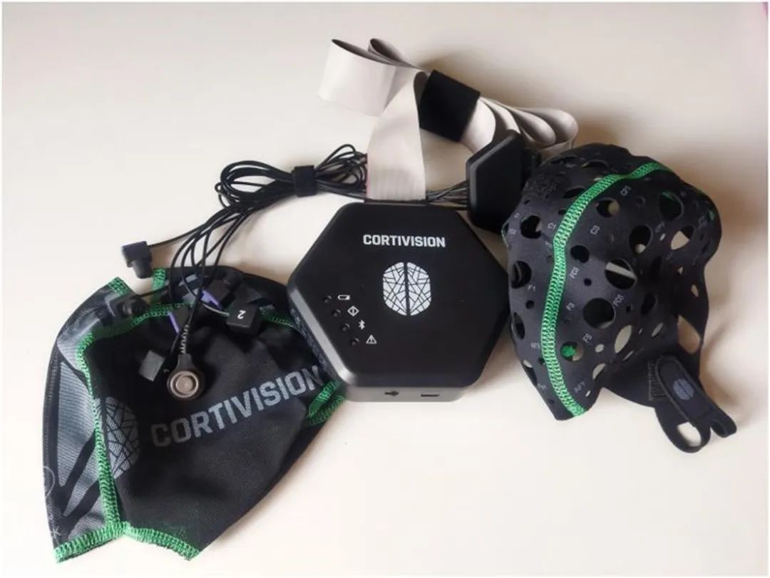


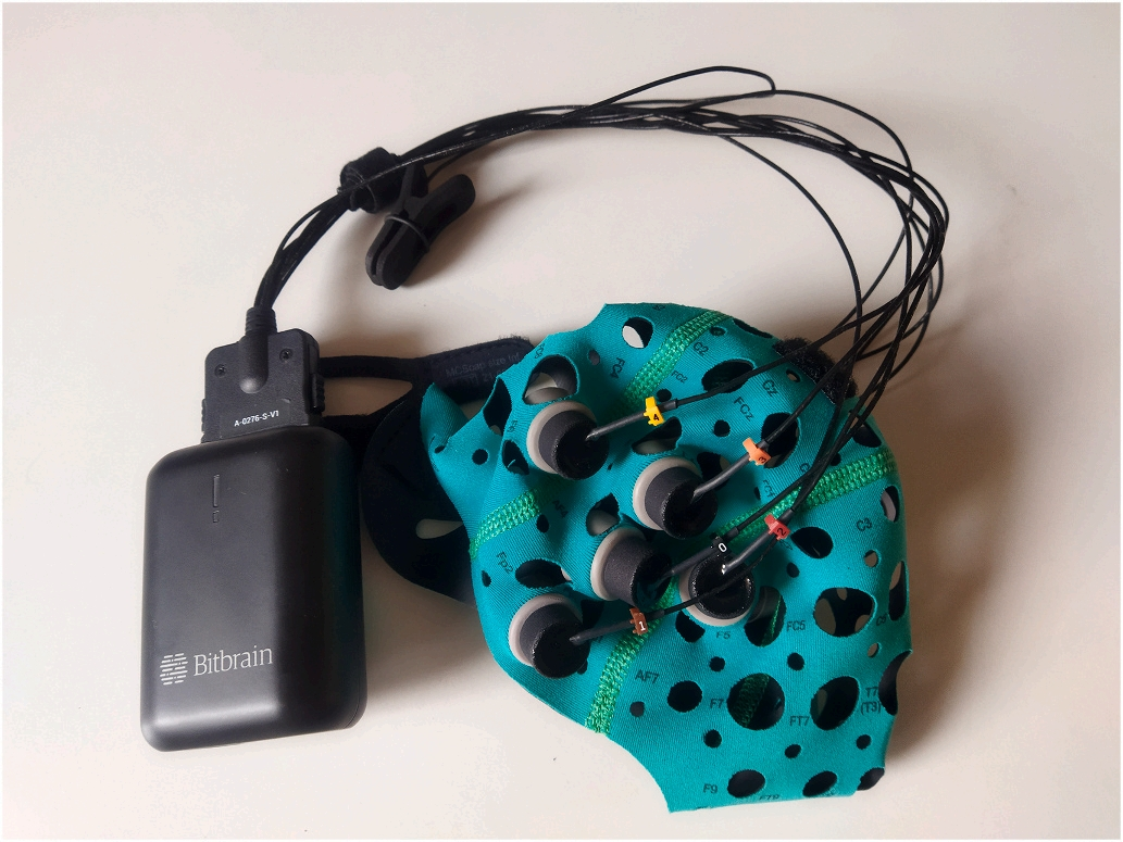


Sociodemographic variables and mother's lifestyle information were collected at the beginning of the study, and participant responses were captured in brain regions throughout the study by using a Cortivision near infrared device combined with an electroencephalography (EEG) device.
04
data analysis


Methods of analysis
Changes in the relative power of the participants' delta frequency with respect to the relative amount of change in oxyhaemoglobin (HBO) across conditions were analyzed using multifactorial analysis of variance (ANOVA), and the efficacy of the different treatments was analyzed using analysis of covariance (ANCOVA), with birth gestational age and birth weight as covariates.


Results
At baseline, preterm infants have lower EEG (delta wave of electrical brain activity) and fNIRS (oxyhemoglobin) values than term infants. Both reflexology and massage therapy can increase delta wave and oxyhemoglobin levels in children.
summarize
Preterm infants may have developmental deficits relative to full-term infants, and early intervention can help to compensate for the developmental deficits of preterm infants. Reflexive movement therapy and massage have been widely used to assess physical and reflexive responses, but there is little evidence of brain mechanisms activated in infants. This study found both techniques to be feasible and safe, however no differences were found between the two.
Original Message
Rocío Llamas-Ramos, Juan Luis Sánchez-González, Jorge Juan Alvarado-Omenat, Vicente Rodríguez-Pérez, Inés Llamas-Ramos.
Brain electrical activity and oxygenation by Reflex Locomotion Therapy and massage in preterm and term infants. a protocol study,
https://doi.org/10.1016/j.neuroimage.2024.120765.
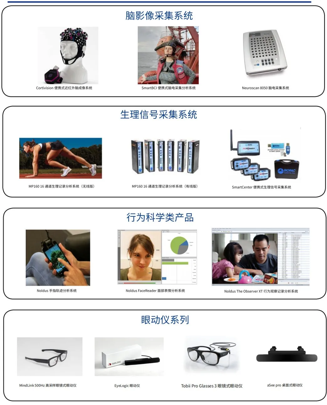
Cortivision Wireless Portable Near Infrared Optical Brain Imaging System
![]()
![]()
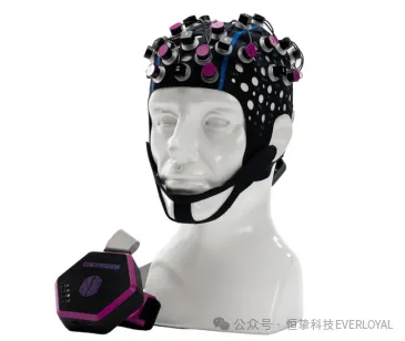

The PHOTON CAP device developed by Cortivision is a wireless portable near-infrared brain optical brain imaging system (fNIRS), which is a device that can effectively measure changes in the concentration of hemoglobin in the brain. The device records and analyzes changes in oxyhemoglobin concentration and deoxyhemoglobin concentration in active brain regions during the perception of external stimuli, and speculates on the interrelationships between perceptually active brain regions and their associated brain regions.
The Cortivision PHOTON CAP is a 37-channel device consisting of 16 transmitters (including 4 shortwave transmitters), 10 detectors. The device is wireless, portable, lightweight, safe and highly compatible, and can be used in conjunction with a wide range of devices such as VR, EEG/ERP, oculomotor, and physiological multidetector.
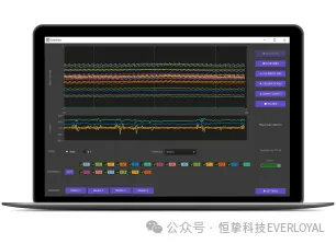


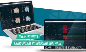


-
Real-time fNIRS and IMU raw data, oxyhemoglobin, deoxyhemoglobin, and total hemoglobin maps; -
No coding is required to go from raw data to results; -
Intuitive graphical user interface; -
Supports .snirf and BIDS formats; -
Has a visualization of the result graph.
Cortivision PHOTON CAP wireless portable near-infrared optical brain imaging system has a wide range of applications and important scientific research significance in the direction of human factors engineering, neuromanagement, neuroengineering management, human-computer interaction, human-computer-environmental systems engineering, environmental behavior, architectural design, and user experience.
Company Profile
