


01
summary
Electroencephalography (EEG) and functional near-infrared spectroscopy (fNIRS) are noninvasive functional neuroimaging techniques. On a single-peak basis, EEG has poor spatial resolution but exhibits high temporal resolution. In contrast, fNIRS provides better spatial resolution. an important advantage shared by EEG and fNIRS is that both modalities have good portability and can be integrated into compatible experimental setups, which provides a good basis for multimodal coupling of EEG-fNIRS. Although an increasing number of studies using synchronized fNIRS-EEG designs have been reported in recent years, the methodology of past studies remains unclear. To fill this knowledge gap, this paper critically summarizes the current state of the art of analytical methods used for simultaneous fNIRS-EEG studies. Finally, the challenges and potential for synchronized fNIRS-EEG data analysis in future studies are highlightedPower.
02
principle
The human brain consists of billions of neurons, each of which forms numerous synapses that create a complex network with trillions of connections, thus enabling our brain to function in an adaptive manner. It is believed that human brain functions and related behaviors are executed by complex neural activations and networks, and that these internal activities generate electrical activity (direct effects) accompanied by hemodynamic responses (indirect effects). Depending on the source of the signal, these brain imaging techniques can be broadly categorized into two groups, the first refers to imaging techniques that reflect neural electrical activity by detecting electrical signals on the scalp surface, the most representative method in this category is (EEG). The second category consists of indirect imaging methods that rely on hemodynamic (cerebral blood flow, cerebral blood volume) and metabolic (glucose and oxygen utilization) responses induced by neural activity, and the common techniques in this category include functional near infrared spectroscopy (fNIRS), functional magnetic resonance imaging (fMRI), and positron tomography (PE). From this perspective, EEG and fNIRS are becoming increasingly popular in the research community and clinical practice due to their unique properties, especially their noninvasiveness, mobility, and flexibility.
2.1 fNIRS Foundation
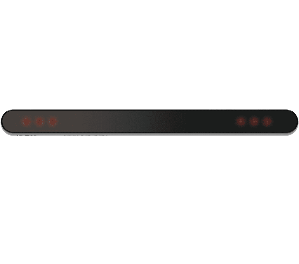
fNIRS is an optical imaging technique used to noninvasively study hemodynamic responses in the brain, typically utilizing near-infrared light of varying wavelengths (between 600 and 1000 nm) to penetrate the scalp and reach the cortical surface, and calculating changes in the concentration of oxygenated hemoglobin (HbO) and deoxygenated hemoglobin (HbR) according to the modified Beer-Lambert law. fNIRS is particularly useful for studying functional activation within the brain due to the intrinsic relationship between neural activity and hemodynamic responses in the brain. The technique is particularly useful for studying functional activation within the brain due to the intrinsic relationship between neural activity and hemodynamic responses in ways such asFigure A.
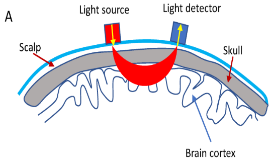
seekA
2.2 EEG Basics
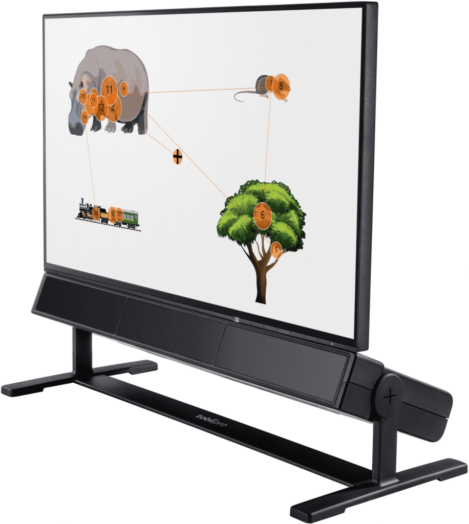
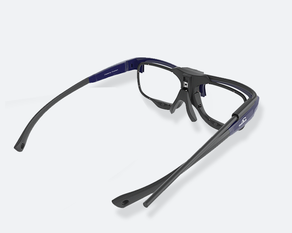
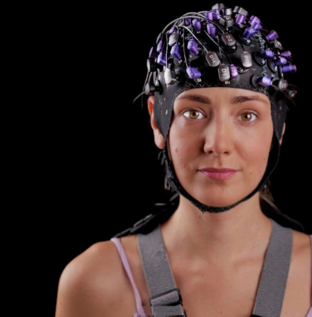
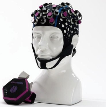
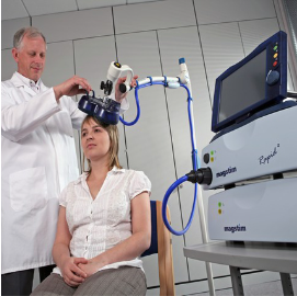
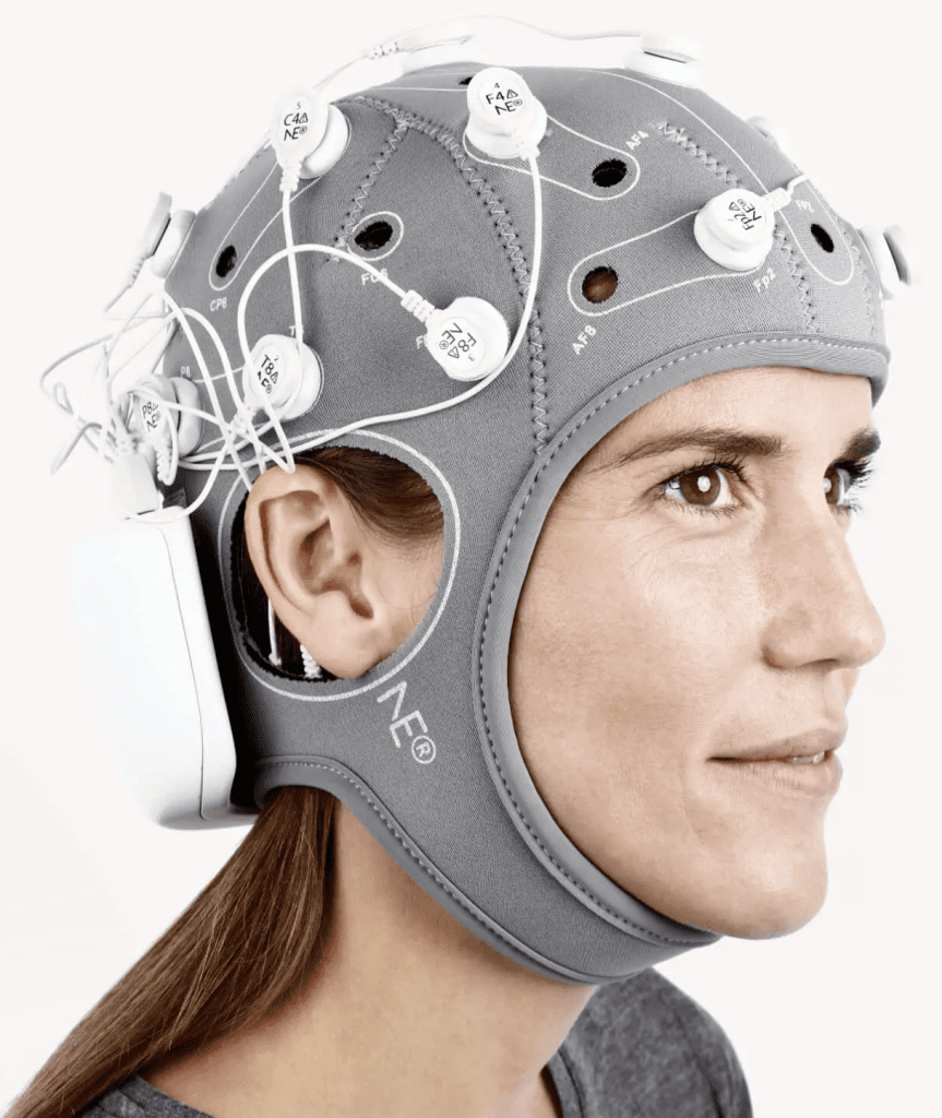
Electroencephalography (EEG) is thought to be primarily due to the synchronization of postsynaptic potentials in cortical pyramidal neurons. That is, when the brain is activated, tens of thousands of pyramidal neurons in the cortex discharge synchronously, where the dendrites of the neurons are oriented, parallel to each other and perpendicular to the cortical surface, thereby inducing sufficient summation and propagation of electrical signals to the scalp. Typically, EEG signals are measured by electrodes placed on the subject's scalp, including a reference (Ref) electrode and a grounding electrode (GND). The recorded EEG signals represent large-scale neural oscillatory activity. Acquisition methods such asFigure B.
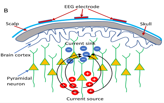
seekB
2.3fNIRSPrinciples and advantages of synchronized integration with EEG
EEG andfNIRSThe basic principle behind the combination also relies on a physiological phenomenon within the brain called neurovascular coupling. Neural activity is inherently accompanied by cerebral blood flow (CBF)ripplesMove, CBFDelivering oxygen and nutrients to neurons. Specifically, when neurons are activated in a specific brain region, blood will flow to that region to meet theneuronsFor glucose and oxygendemand for, which leads to fluctuations in HbO and HbR, which can be measured by fNIRSdetected by the technique. The so-called neurovascular coupling constitutes an integrated fNIRS of brain activity-Theoretical basis of EEG imaginge.g.Figure C.
in order tochange or replaceComprehensive exploration of the functional activity of the brain, compared to unimodal approachesIntegrated fNIRS-EEGmultimodalmethods offer many benefits by utilizing their respective strengths; EEG provides good temporal resolution, theand fNIRSoffersrather or relatively goodspatial resolution, and for the noiseHighly robust. In addition, the EEG and fNIRSsignals are associated with neuronal electrical activity andhemodynamicsresponse correlation, providing built-in validation of the identified activity. Thus, measurements obtained from each of these two modalities provide complementary information related to functional brain activity.
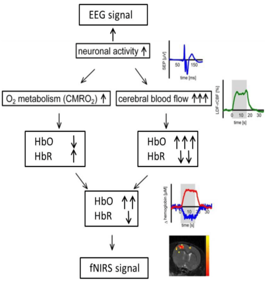
seekC
03
Method
BUSINESS
3.1fNIRSBasic pre-processing of signals
A particularly important step in fNIRS data preprocessing is signal quality checking and artifact correction. fNIRS signal quality can be affected by a variety of confounding noise sources, such as instrumental noise (e.g., due to unstable light sources, electronic noise), physiological disturbances (e.g., respiration, heartbeat), or motion artifacts. Instrumental noise and physiological disturbances are mostly located in the constant frequency range. For example, instrument-induced noise is about 3-5 Hz, and respiration and heartbeat are located at 1-1.5 Hz and 0.2-0.5 Hz, respectively.Therefore, these noises can be minimized by applying a bandpass filter. Motion artifacts in the form of spikes or baseline offsets are a typical noise class in raw fNIRS signals, especially in data collected from groups of children or in experimental tasks that include motion (e.g., walking or talking). Various algorithms have been developed to identify and correct motion artifacts in raw fNIRS signals, such as spline interpolation, wavelet-based methods, or principal component analysis. basic preprocessing procedures for fNIRS data are as followsFigure D.
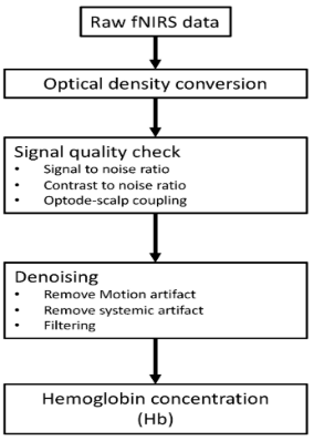
seekD
3.2Basic preprocessing of EEG signals
EEGSignal alsoOften contaminated by different artifacts from internal and external sources. Internal artifacts include physiologic activity of the subject (e.g., electrocardiogram, muscleElectricity, ,blinking) andof the head and of the limbsMotion. External artifacts mainly include environmental/instrumental interference, cable movement,Electrodes not in good contact with scalp. Elimination of internal artifacts relies on additional measurements (e.g., EEG/ECG/accelerometer) or signal decomposition algorithms (e.g., ICA/PCA). External artifacts can be removed by filters, signal decomposition algorithms (e.g., ICA), or artificial segments of theshearing (force)to removeEEGBasic pre-processing of dataprocess asFigure E.
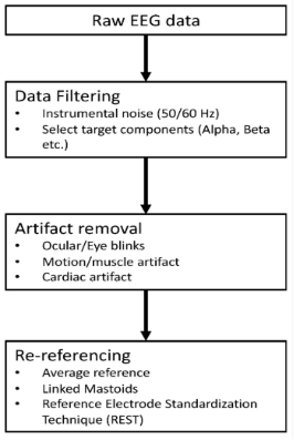
seekE
3.3 EEG-basedfNIRSanalyze
InfNIRS-EEGsynchronizationIn the study, EEG-derived features were used toreinforceFNIRSofanalysis, which is often referred to asfor "based onEEGoffNIRS Analysis".provides a particularly novel and straightforward solution for studying neurovascular coupling. In a typicalfNIRSIn the analysis.fNIRSSignals are typically regressed by a general linear model (GLM), which identifies cortical areas activated by specific stimuli by convolving a typical hemodynamic response function (HRF) with a Boxcar or pulse function representing a consistent temporal profile of the experimental paradigme.g.Figure F.
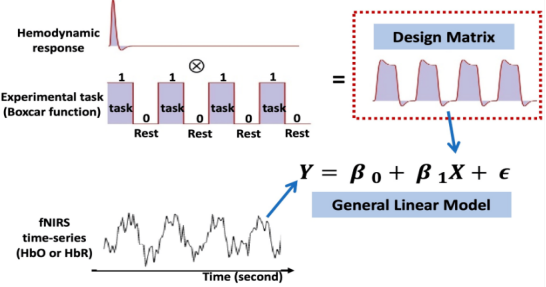
seekF
Common IndependentfNIRSThe main limitation of the analysis is that in real-world settings, neuronal responses to repeated trials or stimuli are time-varying throughout the experiment, which is inconsistent with the boxcar function commonly used in constructing design matrices for GLM analyses.And the EEG-basedfNIRSThe core idea of the analysis is to replace or adjust f with the time or frequency of interest from the EEG signalNIRS Boxcar function in GLM analysis. Based on the linear assumption of neurovascular coupling, the neural activity features extracted from the EEG can be better estimated after the HRF convolutionfNIRSofresponse, thereby increasing the efficiency of recognizing relevant activity regions induced by the experimental task.Figure GsummarizesEEG-basedfNIRSA broad analytical framework for analysis.Table 1summarizesconcerningcarry outEEG-basedfNIRSA study of analysis.
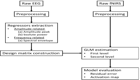
Figure G
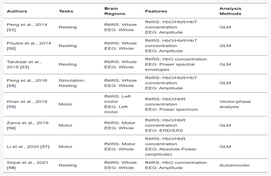
Table 1
3.4 Based on fNIRSEEG analysis of the
EEG due to high time resolution and portabilitybeThe most widely used neuroimaging technique for measuring fast neuronal electrical activity. However, one limitation of scalp EEG is the volume conduction problem; a single electrode on the scalp can be acquired from multiple sources (cortical activity, subcortical activity, external noise, etc.)Electricityactivities, which makes it difficult to accuratelyof determining the source of electrical activity. WhereasfNIRSfavorableofSpatial resolution, mostly based onfNIRSofEEGThe studies have all focused on the use offNIRSof spatial priors to enhance the estimation of EEG source activity.Figure HShows the basic concepts of EEG source imaging and based on thefNIRSof the EEG source imaging analysis process.
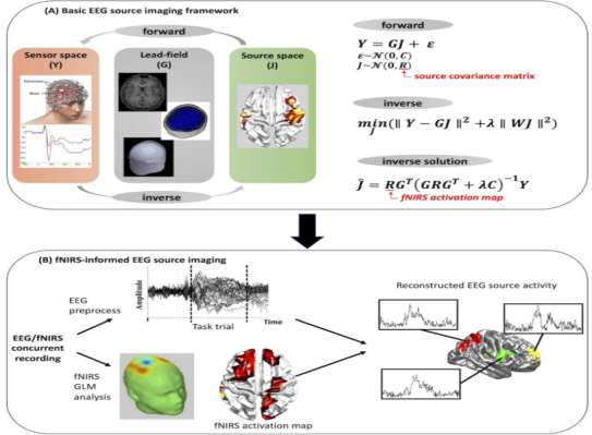
Figure H
3.5EEG-fNIRSParallel analysis of the
The above two sectionsdescribes the relationship between EEG and fNIRSof targeted integration analysis. However, most of the concurrent EEGs available for review-fNIRSStudies have focused on parallel analysis of two complementary techniquesand on research.These studies broadlymayIt is summarized in two categories:(1) Brain-Computer Interface (BCI) (2) Characterization of typical and atypical brain functions.
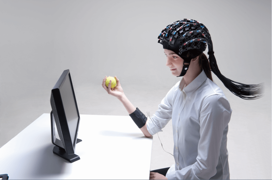
The BCI system was developed to allow users to control computers or external devices based directly on brain activity. Specifically, by fusing features from both modalities, the hybridfNIRS-EEG in various tasks such as motor imagery and execution.Classification and decoding are more accurate than in a single mode.On the other hand.fNIRSand the complementary properties of EEGIt also bringsHemodynamics and electricity of brain activity associated with various functionsphysiological activation modeof a wide range of studies, such as brain load, affective states, and intellectual functioningetc.
Despite the different goals of the two types of studies mentioned above, most of the studies dealing with concurrent fNIRSand EEG data tend to follow similar steps, which mainly include feature extraction, feature fusion and classification. In the review literature, the widely usedfNIRSCharacteristics are usually derived from changes in the concentrations of HbO and HbR, including mean, slope, skewness, kurtosis, peak, variance, and median. Concurrent fNIRS-The typical EEG features used in EEG analysis are highly dependent on the experimental task. In the case of motor tasks, power spectral densities and spatial patterns are widely used, and studies involving cognitive tasks typically employ features related to signal band power.In classification, most of the existing research uses traditional machine learning techniques such as decision trees, linear discriminant analysis (LDA), support vector machines (SVM) and k-nearest neighbor (KNN).
04
fNIRS-EEGconformanalyzeoflimitationswithfuture direction
fNIRSand EEG are portable, noninvasive andRelatively low costof brain imaging, enabling researchers to study brain function under conditions not suitable for other neuroimaging modalities (e.g., fMRI and MEG). Thus, the acquisition and analysis of concurrent, integratedfNIRS-EEG Data May Reveal More Comprehensive Information Related to Brain Activity.even though fNIRSVarious fusion methods of fNIRS and EEG signals allow imaging studies of brain activity using richer information, but most such integrative analyses still rely on data from fNIRSand EEG time series data to extract signals.Neural activity varies over time, so a more dynamic analysis is needed to improve the accuracy of modeling actual brain function. Therefore, exploring with finer temporal resolutionfNIRSand EEG signaling are important for dynamic interactions.
Although there are a number of approaches to concurrentfNIRS-EEG for comprehensive analysis, but most studies have utilized feature-based fusion of these two modalities, such as hybrid BCI systems or feature-basedfNIRS(e.g., mean HbO) and correlation analysis between EEG-based features (e.g., power spectra). Such analyses allow only a coarse characterization of the neurovascular coupling of potential brain activity. With respect to the results from thefNIRS-It remains questionable how the results obtained in the comprehensive EEG analysis reflect the interaction between neuronal electrical activity and the resulting hemodynamic response. Therefore, future work could be conducted on fNIRSand EEG data were performedchange or replacesynthesizeAnd dig deeper.
fNIRSand EEG have helped to link brain imaging techniques in laboratory settings to real-world applications because of their high mobility, non-invasiveness, and low cost. However, few studies have validated the feasibility of using this multimodal approach to meet the needs of multiple real-world scenarios.Therefore, priority objectives for future researchpossibleFocus on improving experimental designData analysis andEcological validity of algorithmsfirst (of multiple parts).
apart fromfNIRSand EEG outside of the methodological integration perspective.the main body of a bookI would also like to emphasize that the instrument is under developmentMatters requiring attention, usingUser-friendly materials for comfortable contactto make light poles.Lightweight integrated design for an enhanced measurement experienceto improve the user experience.
Reference List
Li, R., Yang, D., Fang, F., Hong, K. -S., Reiss, A. L., & Zhang, Y. (2022). Concurrent FNIRS and EEG for Brain Function Investigation: a Systematic, Methodology-Focused Review. Sensors, 22(15), 5865. https://doi.org/10.3390/s22155865
If there is any infringement, please contact us for removal!
Company Profile

Ltd. is an innovative high-tech enterprise focusing on cutting-edge technology, specializing in brain science, neural management, human factors engineering, biomechanics, anthropomorphic environments and XR simulation reality and other multidisciplinary cross-cutting fields. The company is invested by Zhongke (Guangdong) Science Group, relying on the scientific research strength of Guangdong Human Factors Technology Research Institute and Wuhan Human Factors Engineering Technology Research Institute, and has constructed a professional operation system integrating research and development, production, sales and technical service to provide customers with one-stop, high-quality scientific and technological solutions.
With excellent innovation ability, Hengbest Technology has been awarded many invention patents, software copyrights and registered trademarks, selected in many authoritative lists such as National High-tech Enterprises, and participated in the compilation of national standards and group standards. The company has been serving universities and research institutes for a long time, and has cooperated deeply with many national societies such as the Chinese Society of Ergonomics, the Chinese Psychological Society, the Architectural Society of China, etc. The company organizes and participates in more than 40 academic conferences every year to promote technical exchanges and the development of the industry.
With the concept of "Technology Empowerment, Innovation Drive", Hengzhi Technology is committed to become a leading technology enterprise in the industry, to help national scientific and technological progress and social development, and to work together with partners from all walks of life to achieve a better future of technological empowerment.

Scan the code to follow us


