


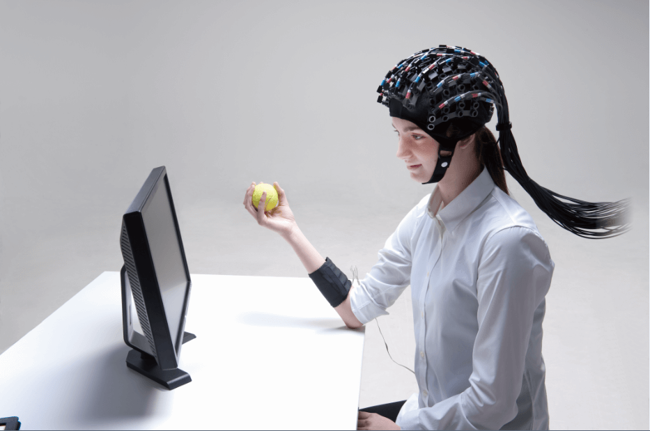


Electromyographic signals (EMG) are bioelectrical signals generated during muscle activity that can be captured by non-invasive surface electrodes. This electrical signal can accurately quantify the degree of muscle activation, fatigue and efficiency of neural control, providing core data for objective assessment of neuromuscular function.
As a well-established electrophysiologic technique, theEMGThe analysis has been widely used in key areas such as clinical rehabilitation diagnosis, sports science performance optimization, human-computer interaction interface development (e.g., prosthetic limb control), bionic manipulation of industrial robots, and aerospace medicine health monitoring, and it is an important cornerstone for connecting physiological mechanisms with engineering applications.
Electromyographic signals (EMG) are essentially the sum of electrical activity generated by muscle fibers in response to nerve stimulation. When the brain sends a command to exercise, the electrical signal is transmitted along the nerve to the muscle, triggering a change in the membrane potential of the muscle fiber, which ultimately triggers a muscle contraction through an "electrochemical switch".
EMGs are categorized into two main types based on the acquisition method:
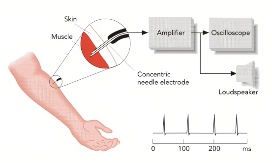
Insertion electromyography (iEMG):Acquisition by direct insertion of needle electrodes into the muscle is high signal quality but invasive.
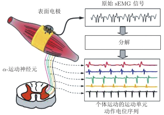
Surface electromyography (sEMG)The EMG signal is collected by electrodes attached to the skin surface, which is non-invasive and convenient, but the signal is susceptible to tissue filtering and noise interference. sEMG signals are extremely weak (0-1.5mV), with the main energy concentrated in the 10-150Hz range. This microvolt-level signal must be precisely amplified and processed to be effectively interpreted, which is the core challenge in the design of EMG acquisition devices.
EMG (Electromyography) records the electrical signals produced by your muscles when they are active, as if they were "listening" to the commands your brain sends to your muscles. When you lift a water bottle, type, or even just get nervous, the brain sends electrical signals through your nerves to your muscles, triggering them to contract, and EMG is the "outward sign" of these electrical signals.
Different EMG changes correspond to different physiological states:
1. Larger amplitude: muscle exertion is increased and more motor units are activated;
2. Decrease in frequency: the muscles begin to fatigue and the electrical signal conduction slows down;
3. Weak or abnormal signals: may reflect impaired nerve conduction or impaired muscle function;
4. change in activation pattern: suggests a change in motor control strategy or coordination;
EMG is an "electrical activity map" that connects brain-nerves-muscles, revealing not only how we exert force, but also whether the body is stressed, fatigued, or in an abnormal state.
1. Principle of measurement:
differential amplifierGreat:The muscle potential difference is collected through two (+/-) surface electrodes and then, together with the ground electrode, fed into a differential amplifier to improve the signal-to-noise ratio and suppress common mode interference such as industrial frequency.
Bandpass/trap filtering:Band-pass filtering of the amplified analog signal (Typical 20-450 Hz) to remove low-frequency drift and high-frequency noise, with optional 50/60 Hz Trap filtering to further remove industrial frequency interference.
Analog-to-digital conversion (ADC):The filtered analog EMG signal will be displayed as ≥1 kHz The sampling rate and ≥16 bit(or similar) resolution digitization for easy subsequent processing and storage.
These three components - differential amplification, filtering, and digitization - are at the heart of all sEMG acquisition systems and together determine signal quality and measurement reliability.
2. Measurement methods and equipment
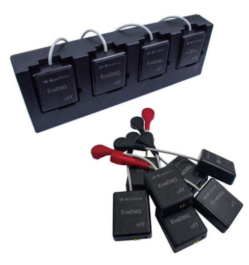
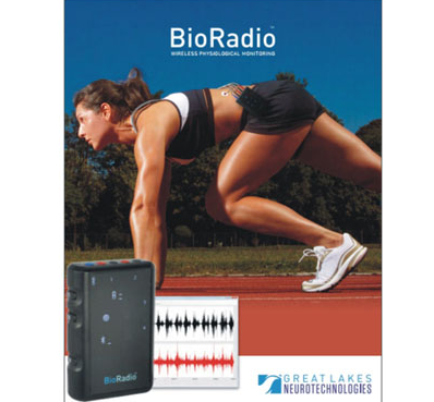
(1) Bioradio
Two electrodes were placed at the fullest part of the target muscle belly, spaced approximately 2-3 cm; grounding electrodes were applied to the bony prominences.
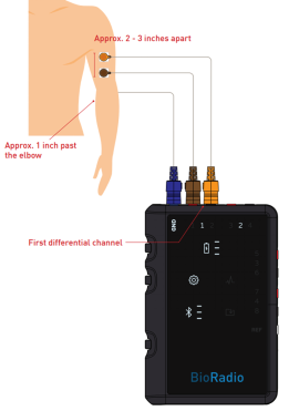
BioRadio is suitable for mobile, wearable and long-term monitoring with wireless transmission and local recording.
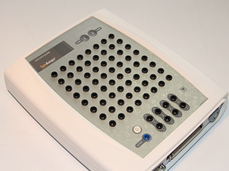
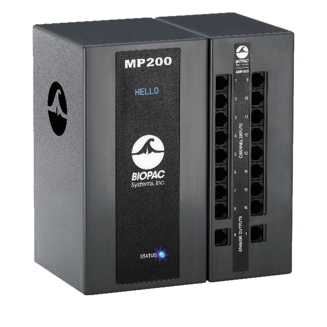
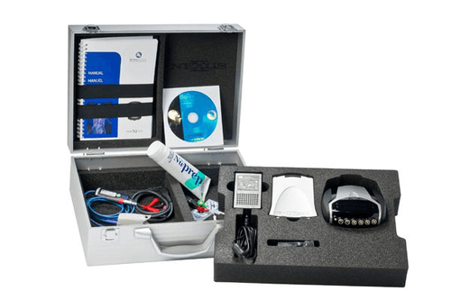
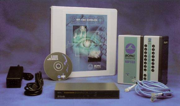
(2) Biopac
Any one of the muscles measured was selected and the electrodes were connected to two thirds of the muscle at both ends.
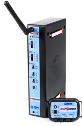
Biopac, on the other hand, is known for its powerful amplification, filtering, and analysis software, and is better suited for high-precision, reproducible research or teaching scenarios in the lab.
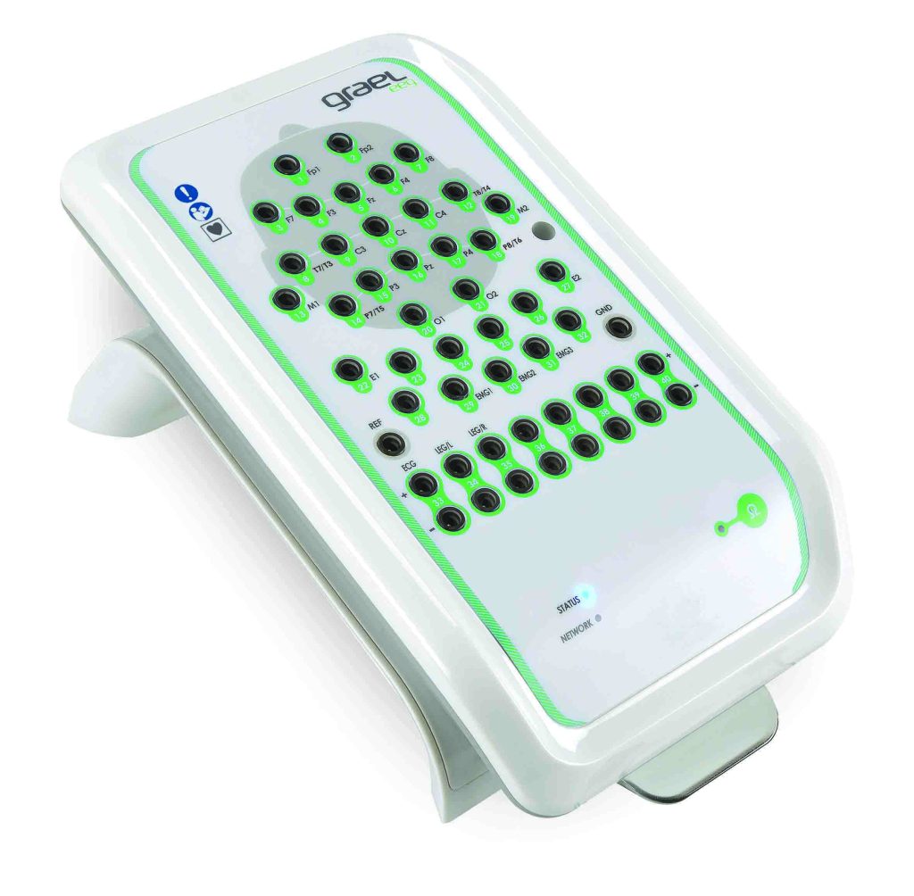
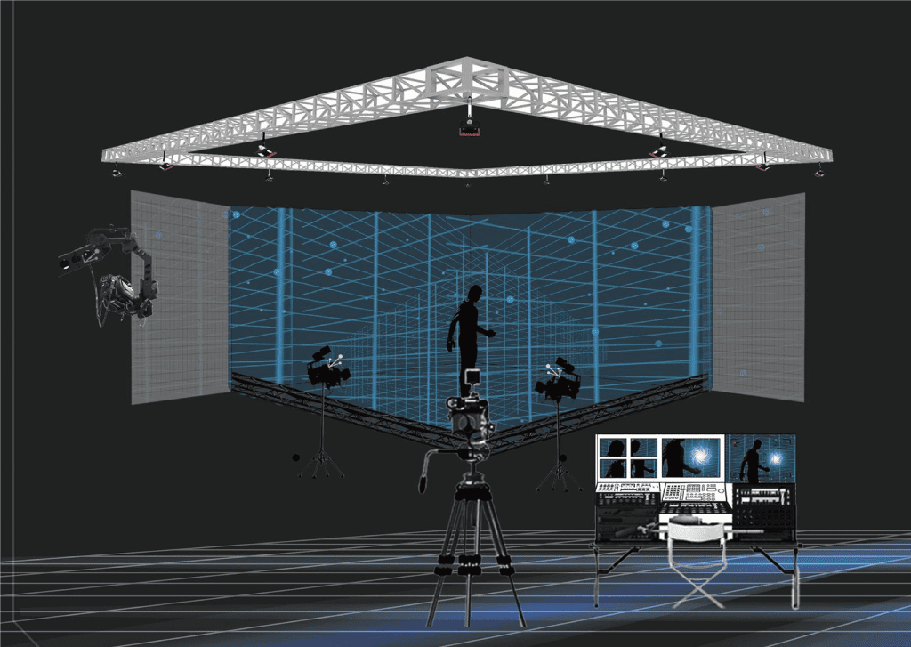
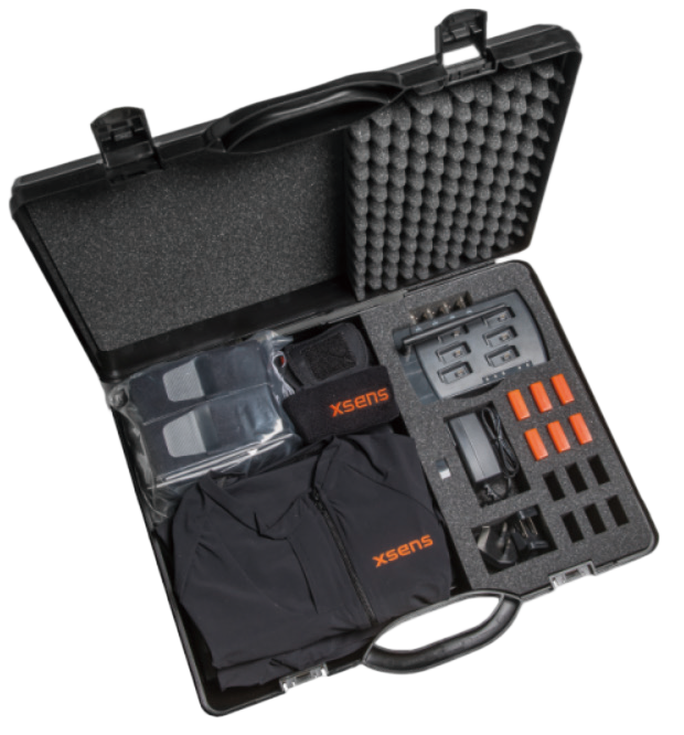
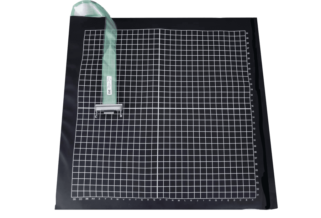
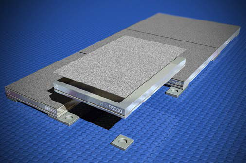
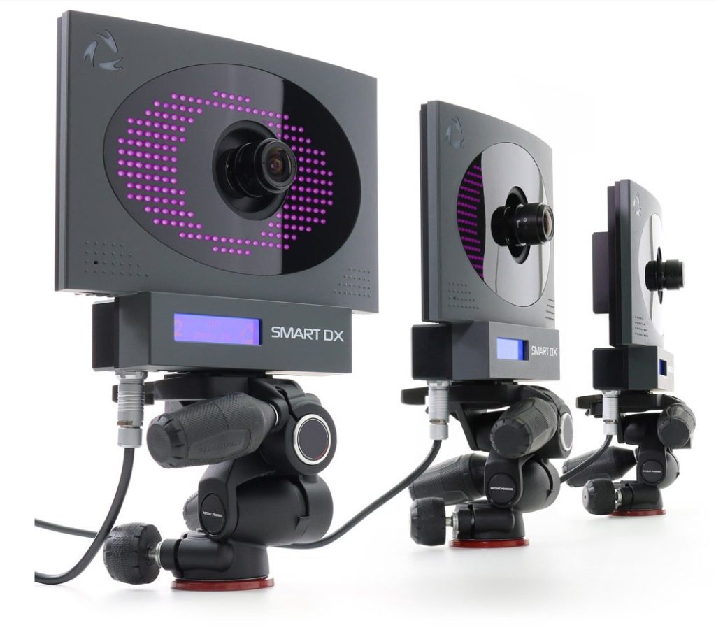
(3) BTS
The EMG probe should be placed in the middle of the muscle of interest, avoiding tendon attachment points. The electrodes must be aligned parallel to the muscle fibers.
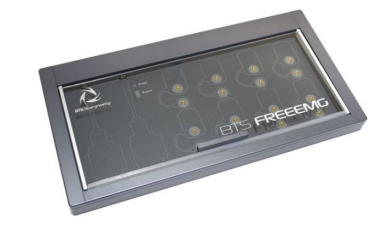
BTS wireless transmission, free choice of indoor and outdoor environment, suitable for mobile monitoring.
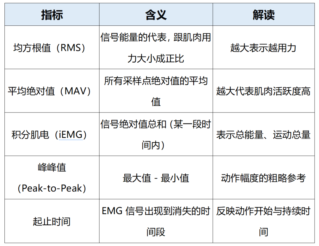
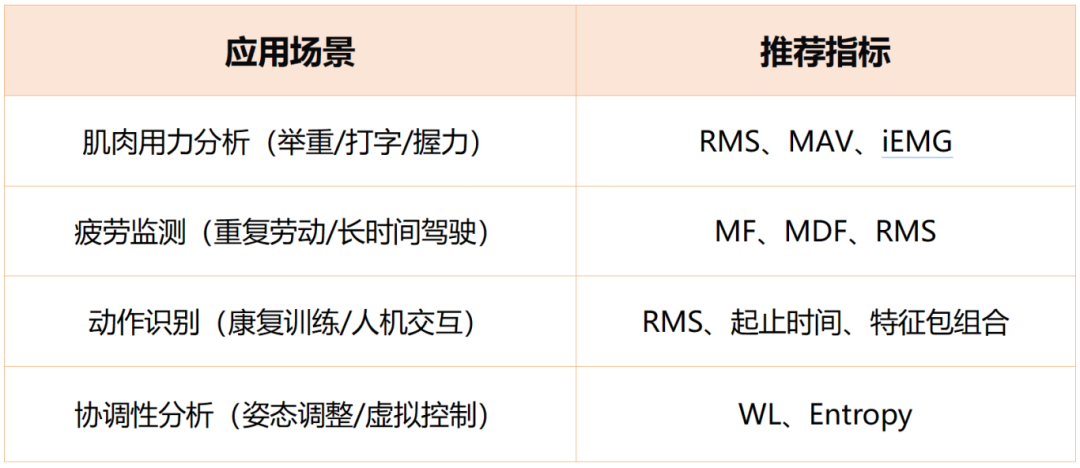
Generally, when analyzing EMG signals, the two main types of metrics are focused on the time domain and the frequency domain:
time domain index (TDI)
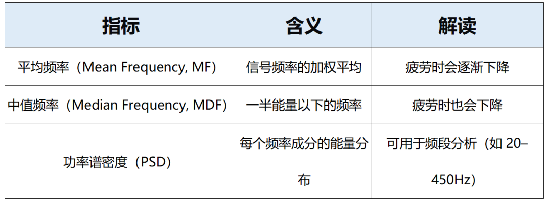
frequency domain index
- EMG amplitude/RMS is considered as a measure of strength output, when EMG amplitude or RMS level is high, it means more motor units are mobilized, reflecting high strength output; conversely, when EMG amplitude or RMS level is low, it reflects low strength output. Meanwhile, when the RMS level rises and then falls or the amplitude continues to fall, it indicates the accumulation of fatigue and a decrease in muscle output; conversely, when the RMS is stable or maintains its level continuously, it reflects a non-fatigued muscle state.

- The magnitude of explosive force can be reflected by the rate of rise of EMG (RFD_EMG). When the rate of rise of EMG is fast, the neuromuscular system is able to rapidly mobilize a large number of motor units in a very short period of time, reflecting high explosive force; conversely, when the rate of rise of EMG is slow, reflecting low explosive force.

Important measures of muscle fatigue include theMedian frequency (MF) / Mean Frequency (MNF) when theMF or MNF levels are low, the high-frequency component is attenuated, reflecting high muscle fatigue; conversely, high MF/MNF levels reflect low muscle fatigue.

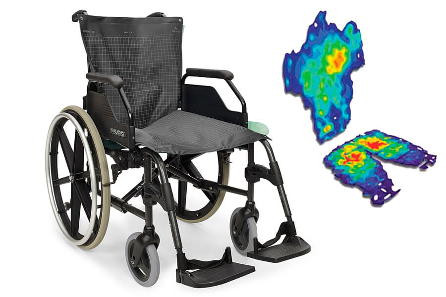
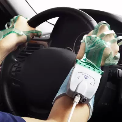
- Rehabilitation training process.Mean Absolute Value (MAV)It's an observation.muscle activation modeOne of the measures that reflect the corresponding movement category (e.g., bending, reaching, or grasping) is when MAV levels show a specific spatial distribution between multiple channels; conversely, when MAV levels do not differ significantly, they reflect a resting or no-movement state.

Company Profile

With excellent innovation ability, Hengbest Technology has been awarded many invention patents, software copyrights and registered trademarks, selected in many authoritative lists such as National High-tech Enterprises, and participated in the compilation of national standards and group standards. The company has been serving universities and research institutes for a long time, and has cooperated deeply with many national societies such as the Chinese Society of Ergonomics, the Chinese Psychological Society, the Architectural Society of China, etc. The company organizes and participates in more than 40 academic conferences every year to promote technical exchanges and the development of the industry.
恒挚 Technology upholds the concept of "doing our part for the cause of scientific research", and is committed to becoming a leading scientific research-supporting science and technology enterprise, contributing to the progress of national science and technology and social development, and joining hands with partners from all walks of life to achieve a better future empowered by science and technology.

Scan the code to follow us


